货号: CL-0074
Price: ¥1600 ¥1600
生长培养基: RPMI-1640[PM150110]+10% FBS[164210]+1% P/S[PB180120]
产品信息
| 产品名称 | DLD-1(人结直肠腺癌上皮细胞) |
| 别称 | DLD 1; DLD1; CoCL3 |
| 生长特性 | 贴壁细胞 |
| 细胞形态 | 上皮细胞样 |
| 冻存条件 |
冻存液:55% 基础培养基+40% FBS+5% DMSO 温度:液氮 |
| 培养方案A(默认) |
生长培养基:RPMI-1640[PM150110]+10% FBS[164210]+1% P/S[PB180120] 培养条件: 气相:空气,95%;CO₂,5%, 温度:37℃ |
| 培养方案B(可选) |
生长培养基: 培养条件: 气相:, 温度:37℃ |
| 推荐传代比例 | 1:3-1:8 |
| 推荐换液频率 | 2-3次/周 |
参考资料(来源文献)
| 背景描述 | DLD-1细胞是由D·L·Dexter和其同事于1977-1979年分离的两株结直肠腺癌细胞株中的一株。在ATCC和其它地方进行的DNA指纹鉴定和染色体组型分析表明DLD-1细胞与HCT-15细胞相似,说明这两者是来自同一个人的不同克隆。DLD-1细胞和HCT-15细胞的遗传起源可通过DNA指纹鉴定证实,但染色体组型分析显示它们缺乏染色体标记一致改变或数目上一致改变。DLD-1细胞的CSAp阴性(CSAp-),p53抗原表达呈阳性(p53抗原产生了一个C→T点突变导致241位的Ser→Phe)。DLD-1细胞角蛋白免疫过氧化物酶染色阳性,癌基因c-myc、K-ras、H-ras、N-ras、myb、sis和fos的表达呈阳性,癌基因N-myc的表达未做检测。DLD-1细胞表达肿瘤特异性核基质蛋白CC-2、CC-3、CC-4、CC-5和CC-6。1979年提交到ATCC的DLD-1细胞代数不明且污染了支原体,其后经过12周多种抗生素联合培养处理,处理之后每周用Hoechst染色和标准培养法检测。其后连续11个月不加抗生素培养,DLD-1细胞所有的检测呈阴性。 |
| 年龄(性别) | Adult Male |
| 组织来源 | 结直肠腺癌上皮细胞 |
| 细胞类型 | 肿瘤细胞 |
| 肿瘤类型 | 肠癌细胞 |
| 生物安全等级 | BSL-1 |
| 致瘤性 | Yes, in nude mice (Tumors developed within 21 days at 100% frequency (5/5) in nude mice inoculated subcutaneously with 1×10<sup>7</sup> cells). |
STR位点信息

STR鉴定图
-
HSPA5 repressed ferroptosis to promote colorectal cancer development by maintaining GPX4 stability(2022)
作者:Ronglin Wang, Lei Hua, Peixiang Ma, Yang Song, Jie Min, Yongdong Guo, Cheng Yang, Junqiang Li, Haichuan Su
期刊:NEOPLASMA
DOI:10.4149/neo_2022_220331N363
影响因子:3.4
引用产品: DLD-1 细胞, SW480 [SW-480] 细胞
-
CAMK2N1 has a cancer-suppressive function in colorectal carcinoma via effects on the Wnt/β-catenin pathway(2022)
作者:Xiaolong Zhang, Lifei Tian, Zeyu Li, Ruiting Liu, Jiao Yu, Bo Liu
期刊:BIOCHEMICAL AND BIOPHYSICAL RESEARCH COMMUNICATIONS
DOI:10.1016/j.bbrc.2022.08.036
影响因子:3.3
引用产品: LS180 细胞, SW480 [SW-480] 细胞, HT-29 细胞, HCT 116 细胞, DLD-1 细胞
-
Circ_KIAA1199 inhibits MSI1 degradation by targeting miR-34c-5p to drive the malignant cell behaviors and tumor growth of colorectal cancer(2022)
作者:Yanbo Zhang, Hailong Yu, Zhen Guo
期刊:Anti-Cancer Drugs
DOI:10.1097/CAD.0000000000001164
影响因子:2.3
引用产品: HCT 116 细胞, DLD-1 细胞
-
Autophagy Induction by Trichodermic Acid Attenuates Endoplasmic Reticulum Stress-Mediated Apoptosis in Colon Cancer Cells(2021)
作者:Junyan Qu, Cheng Zeng, Tingting Zou
期刊:International Journal Of Molecular Sciences
影响因子:5.6
引用产品: HCT 116 细胞, DLD-1 细胞
-
Raltitrexed as a synergistic hyperthermia chemotherapy drug screened in patient-derived colorectal cancer organoids(2021)
作者:Lisi Zeng, Quanxing Liao, Haoran Zhao
期刊:Cancer Biology & Medicine
DOI:10.20892/j.issn.2095-3941.2020.0566
影响因子:5.5
引用产品: SW620 细胞, SW480 [SW-480] 细胞, DLD-1 细胞, COLO 205 细胞
-
Cullin-4B promotes cell proliferation and invasion through inactivation of p53 signaling pathway in colorectal cancer(2021)
作者:Min Zhong, Ling Zhou, Jianping Zou
期刊:Pathology Research And Practice
影响因子:2.8
-
The combination of lactoferrin and linolenic acid inhibits colorectal tumor growth through activating AMPK/JNK-related apoptosis pathway(2021)
作者:Qianqian Yao, Huiying Li, Linlin Fan
期刊:PeerJ
影响因子:2.7
引用产品: Hep G2 细胞, AGS 细胞, HCT 116 细胞, DLD-1 细胞, HT-29 细胞
-
Ubiquitination-mediated degradation of SIRT1 by SMURF2 suppresses CRC cell proliferation and tumorigenesis(2020)
作者:Le Yu, Ling Dong, Hui Li
期刊:Oncogene
影响因子:8.0
引用产品: 293T [HEK-293T] 细胞, HCT 116 细胞, DLD-1 细胞
-
miR-145-5p restrained cell growth, invasion, migration and tumorigenesis via modulating RHBDD1 in colorectal cancer via the EGFR-associated signaling pathway(2019)
作者:Ying Niu, Jinping Zhang, Yalin Tong
期刊:International Journal Of Biochemistry & Cell Biology
DOI:10.1016/j.biocel.2019.105641
影响因子:4.0
引用产品: Caco-2 细胞, DLD-1 细胞, LoVo 细胞, SW480 [SW-480] 细胞, T84 细胞
-
Q1:可以做基因编辑的肠上皮细胞有推荐的吗?
文献显示DLD-1和IPEC-J2两款可做基因编辑: https://www.nature.com/articles/s41598-020-76033-1 https://pubmed.ncbi.nlm.nih.gov/33482281/
-
Q2:为什么我看的文献里的细胞培养条件和你们官网的培养条件不一样呢?
部分细胞是会出现多种培养条件的,我们公司优先选择引种来源的培养条件以及建系者所用培养条件,出现差异的原因是不同实验室在保藏过程中更改了细胞的培养条件,为了避免细胞突然更换培养条件后不适应,建议您优先使用厂家推荐的培养条件培养。
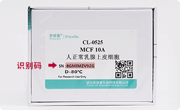
识别码示意图


